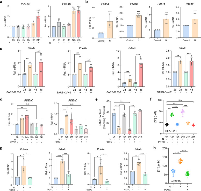
Fig. 5.
SARS-CoV-2 N protein contributed to the sustained elevation in [Cl−]i via NF-κB-PDE4-cAMP signaling pathway in RECs. a The mRNA expressions of PDE4C and PDE4D in the N protein (50 μg/ml)-stimulated BEAS-2B cells (n = 3). b The mRNA expressions of Pde4 in the lung tissues of N protein (0.25 mg/kg)-stimulated mice (n = 3). c The mRNA expressions of Pde4 in the lung tissues of SARS-CoV-2 (1 × 10⁵ PFU)-infected hACE2-transduced mice (n = 3). d The effect of PDTC (100 μM) on PDE4 mRNA expression in BEAS-2B cells stimulated with N protein (50 μg/ml) (n = 3). e The effect of PDTC (100 μM) on intracellular cAMP levels in BEAS-2B cells stimulated with N protein (50 μg/ml) (n = 3). f The effect of PDTC (100 μM) on [Cl−]i of BEAS-2B cells stimulated with the N protein (50 μg/ml) (n = 12 cells). g The effect of PDTC (100 mg/kg) on Pde4 mRNA expression in the lung tissues of N protein (0.25 mg/kg)-stimulated mice (n = 3). h The effect of PDTC (100 mg/kg) on [Cl−]i of mPAECs from mice after N protein (0.25 mg/kg) stimulation (n = 20 cells). Data were shown as mean ± SD, *P < 0.05, **P < 0.01, ***P < 0.001 compared with the control group or indicated by lines, n.s. P > 0.05

