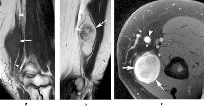
Figure 12.
A 62-year-old female presenting with a swelling in the medial distal arm. (a) Coronal T1W TSE, (b) sagittal T2W FSE and (c) axial fat-suppressed PDW FSE MR images show a lobular homogeneous T1 isointense and heterogeneous T2 hyperintense oval mass (arrows) arising in relation to the ulnar nerve (arrowheads-a). The lesion demonstrates a “target” sign’, with low central T2 signal (arrowhead-b) and is separated from the basilic vein (arrowhead-c) consistent with its extra nodal origin. Histologically confirmed schwannoma.

