Figure 1.

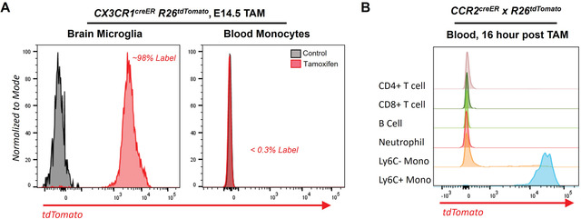
Flow cytometry analysis of fate‐mapped monocytes and macrophages.
A. CX3CR1creER R26tdTomato reporter mice were labeled in utero, embryonic day 14.5 (E14.5), by tamoxifen gavage of pregnant mothers. Labeled pups were weaned and then sacrificed at 8 weeks of age and assessed for labeling efficiency in brain microglia and blood. (Left panel) Brain microglia (CD45+ CD11b+ CD64+, Red line) were labeled with >98% efficiency from embryonic exposure to tamoxifen, whereas unlabeled age‐matched mice showed no labeling (control, Gray line). (Right panel) In the same mice, blood at 8 weeks of age showed no labeling of blood monocytes (CD45+ CD11b+ Ly6G‐ CD115+) in E14.5 labeled (Red) or control (Gray).
B. CCR2creER R26tdTomato reporter mice were gavaged with a single dose of tamoxifen, and labeling efficiency in the blood was analyzed 16 hr later. T cells (CD45+ TCRβ+), B Cells (CD45+ CD19+), Neutrophil (CD45+ CD11b+ Ly6G+), Ly6C‐ Monocytes (CD45+ CD11b+ Ly6G‐ CD115+ Ly6C‐), and Ly6C+ Monocytes (CD45+ CD11b+ Ly6G‐ CD115+ Ly6C+) were assessed for tdTomato labeling, where Ly6C monocytes were efficiently labeled.
