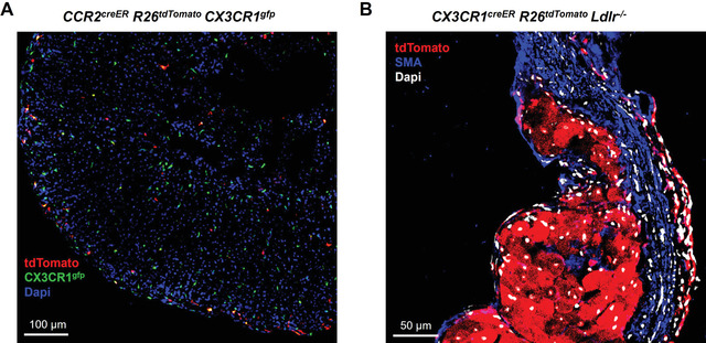
Figure 2.

Fluorescent imaging analysis of fate‐mapped macrophages in tissues.
A. Dual reporter CCR2creER R26tdTomato CX3CR1gfp mice were given a single gavage of tamoxifen (200 mg/kg) and then sacrificed 48 hr later. Adrenal glands were cryosectioned and assessed by confocal microscopy for tdTomato (red) and GFP (green) expression to determine the distribution of differentially expressing cells. Nuclei are labeled with Dapi staining (blue).
B. CX3CR1creER R26tdTomato Ldlr‐/‐ mice were continuously fed a tamoxifen‐enriched high fat diet to induce atherosclerosis while labeling all CX3CR1‐expressing monocytes and macrophages throughout disease progression. After 8 weeks, hearts were collected, fixed in PFA/sucrose, and cryosectioned. Samples were immunostained for smooth muscle actin (SMA), then imaged by confocal microscopy for SMA (blue), tdTomato (red), and DAPI (white) for nuclei.
