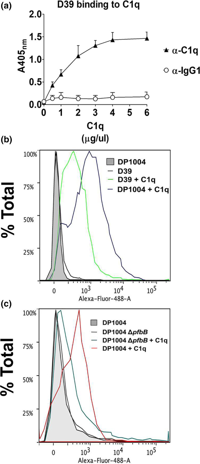
FIGURE 4.

Role of PfbB in pneumococcal binding to C1q. The binding of C1q to pneumococci was measured by ELISA (a) or immunofluorescence flow cytometry (b and c). (a) Multi‐well plates sensitized with pneumococci (5 × 10⁷ CFU/ml) were incubated with increasing concentrations of C1q. Bound C1q was detected using anti‐C1q antibodies (α‐C1q). IgG1, isotype control. Shown are means ± SDs of three experiments performed in triplicate. (b) Binding of C1q (5 μg/ml) to encapsulated D39 (green line) and unencapsulated DP1004 (purple) strains as detected by immunofluorescence flow cytometry using an Alexa‐Fluor‐488‐labeled antibody. (c) Binding of C1q (5 μg/ml) to unencapsulated DP1004 (red) and its isogenic pfbB deletion mutant (ΔpfbB‐DP, dark green), as detected by immunofluorescence flow cytometry using an Alexa‐Fluor‐488‐labeled antibody.
