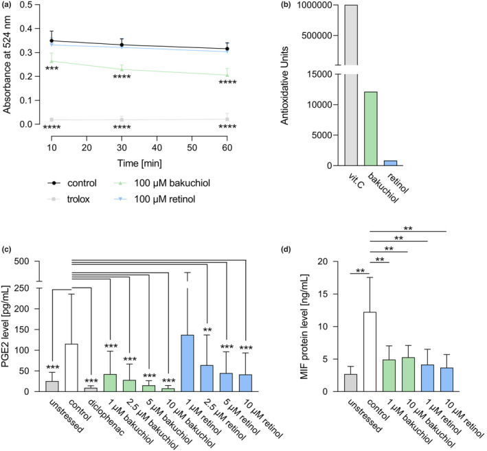
FIGURE 2.

Antioxidative and anti‐inflammatory capacity of bakuchiol and retinol. (a) Antioxidative efficacy of bakuchiol (100 μM) or retinol (100 μM) compared to the high standard trolox (25 μM) and the solvent control determined by a DPPH antioxidant assay through absorbance measurement at 524 nm after 10, 30 and 60 min. N = 12. (b) Antioxidative power expressed in antioxidative units of bakuchiol, retinol or the high standard vitamin C (vit. C) determined using electron spin resonance spectroscopy. (c) ELISA‐based measurement of prostaglandin E2 (PGE2) levels in unstressed human dermal fibroblasts (HDFs), LPS‐stressed control HDFs, and in LPS‐stressed HDFs treated with the high standard diclofenac (25 ng/mL), bakuchiol or retinol (both applied at 1.25, 2.5, 5, 10 μM) for 24 h. N = 12. (d) Macrophage migration inhibitory factor (MIF) protein levels in unstressed HDFs, in HDFs stressed by a DPBS incubation and in stressed HDFs treated with bakuchiol or retinol (both applied at 1 and 10 μM) for 48 h determined by ELISA. N = 10. Results are depicted as mean ± SD. Statistics were performed by RM‐ANOVA with post‐hoc pairwise comparison based on Blom‐transformed ranks for Figure 2a or by a pairwise Wilcoxon signed rank test for Figure 2c and d. Significant differences are marked with an asterisk (**p ≤ 0.01, ***p ≤ 0.001, ****p ≤ 0.0001) [Colour figure can be viewed at wileyonlinelibrary.com]
