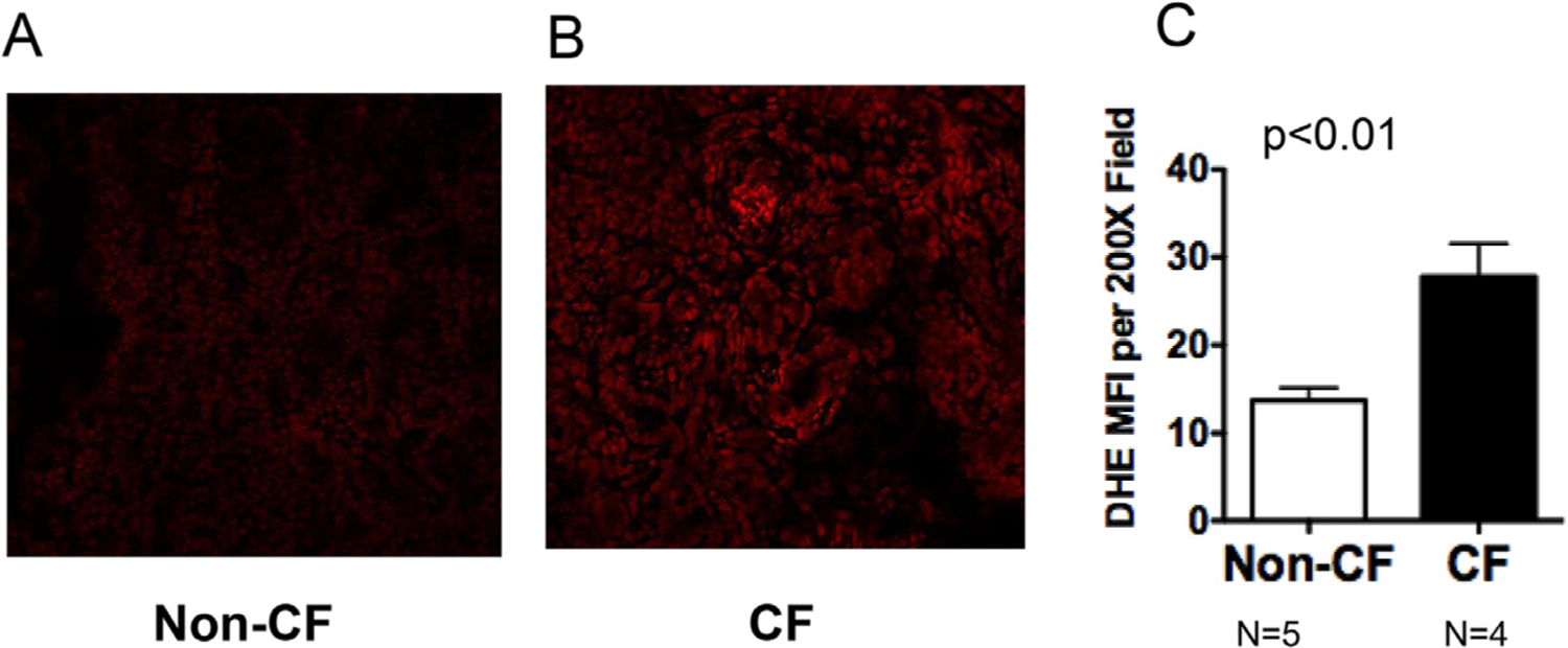
Fig. 2.

CF pancreas shows increased levels of ROS in situ. Pig pancreata were harvested within 12 h after birth and immediately frozen in OCT compound. Samples were cryo-sectioned onto microscope slides and stained with 5 μM dihydroethidium for 30 min at 37 °C. At least three images per tissue section (average 4 sections per tissue) were captured at 200x and 630x using a Zeiss 710 confocal microscope. Image fluorescence intensity was quantified using Image J. Pictures shown in (A) and (B) are representative of quantified data shown in (C). P value is shown.
