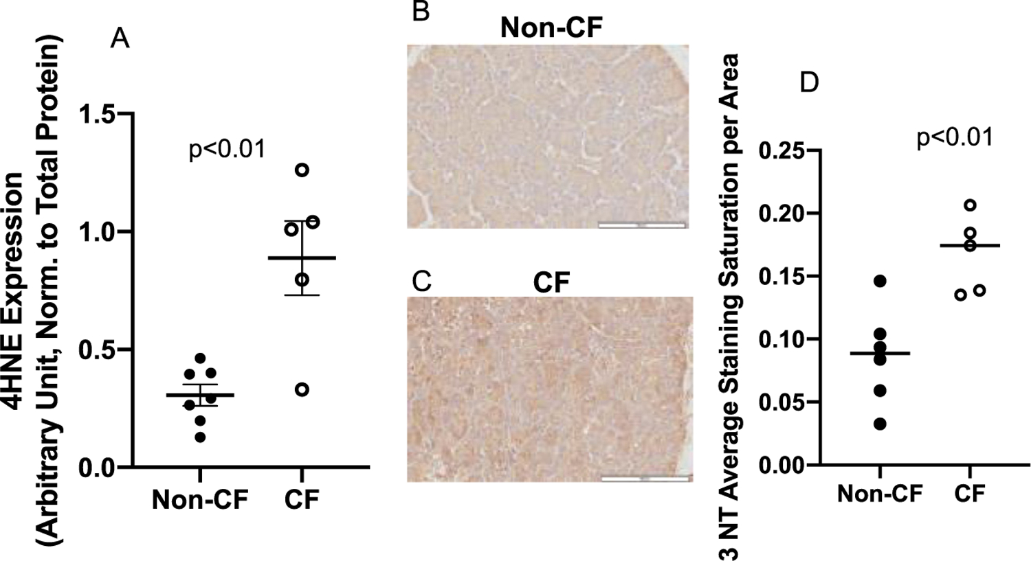
Fig. 4.

4-Hydroxynonenal modified proteins and 3-Nitrotyrosine as determined by immuno-blotting in pancreatic tissue. (A) Pancreatic tissue from pigs with non-CF (N = 7), or CF (N = 5) were probed for 4-HNE expression using immuno-slot-blotting. 4-HNE was significantly elevated in CF pig pancreas relative to non-CF (p<0.01). Data shown were the average staining of samples from three separate blots normalized to total protein via ponceau staining and reported with SEM. Micrographs of pancreatic tissue from (B) non-CF (N = 5) and (C) CF (N = 4) pigs probed for 3-NT-modified protein using immunohistochemistry and (D) quantified according to staining intensity per area. CF pig pancreas displayed significantly increased (p < 0.01) 3-NT staining throughout the pancreas. Data shown are the average of at least 6 sites selected randomly from each tissue section and reported with SD.
