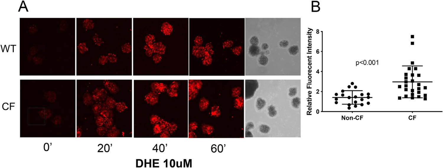
Fig. 5.

CF Islets have increased levels of ROS. (A) Islets were isolated and cultured in glass bottom dishes overnight at 37 °C, 5% CO2. The images were acquired immediately after addition of 10 μM of DHE at room temperature (about 25 °C) without additional CO2, at excitation/emission wavelength of 405/570 nm, using a Zeiss 710 confocal microscope. Images for WT and CF islets were obtained at the exact same gain setting on the microscope. (B) The fluorescent intensity of the images was quantified using ImageJ. Data shown are representative of three separate experiments.
