Figure 1.

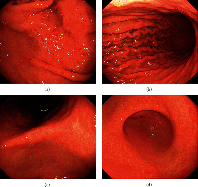
Esophagogastroduodenoscopy images. Endoscopy reveals spotty redness at the gastric fornix (a), mucosal swelling with diffuse redness in the corpus (b), and mucosal atrophy in the gastric angle (c) and antrum (d).

Esophagogastroduodenoscopy images. Endoscopy reveals spotty redness at the gastric fornix (a), mucosal swelling with diffuse redness in the corpus (b), and mucosal atrophy in the gastric angle (c) and antrum (d).