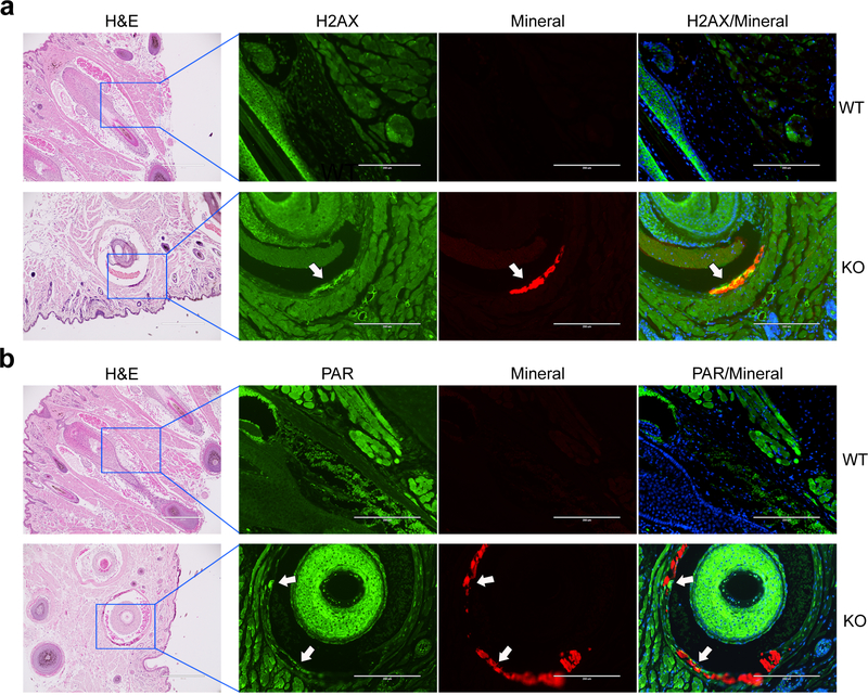
Figure 1. Immunofluorescent staining of H2AX and PAR shows the presence of DNA damage response and PAR deposition in the muzzle skin.
Paraffin sections of muzzle skin from 5 wild-type and 5 Abcc6−/− mice were analyzed. Bar = 200 μm. Arrows indicate the positivity of the green fluorescent signals of H2AX (a) and PAR (b) in association with mineral deposits (red fluorescence), as shown as yellow in the overlay. Cell nuclei were stained with DAPI. Adjacent slides were stained with hematoxylin and eosin for mineral deposits. WT, wild-type; KO, Abcc6−/−.

