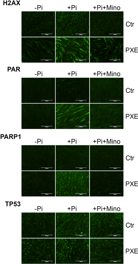
Figure 4. Immunofluorescent staining of H2AX, PAR, PARP1, and TP53 in cultured dermal fibroblasts.
Dermal fibroblasts from three healthy controls and three patients with PXE were cultured in DMEM medium containing 10% fetal bovine serum. Immunofluorescent staining (green fluorescence) was performed in cells cultured in normal medium and medium supplemented with 4 mM Pi with or without 3 μM minocycline for 4 days. The cellular localization of H2AX, PAR, PARP1, and TP53 in cells cultured in medium containing 4 mM Pi was provided in Supplementary Figure S1. Ctr, control; Mino, minocycline; Pi, inorganic phosphate; Bar = 200 μm.

