
Figure 5. Recovery from Anesthetic Inhibition.
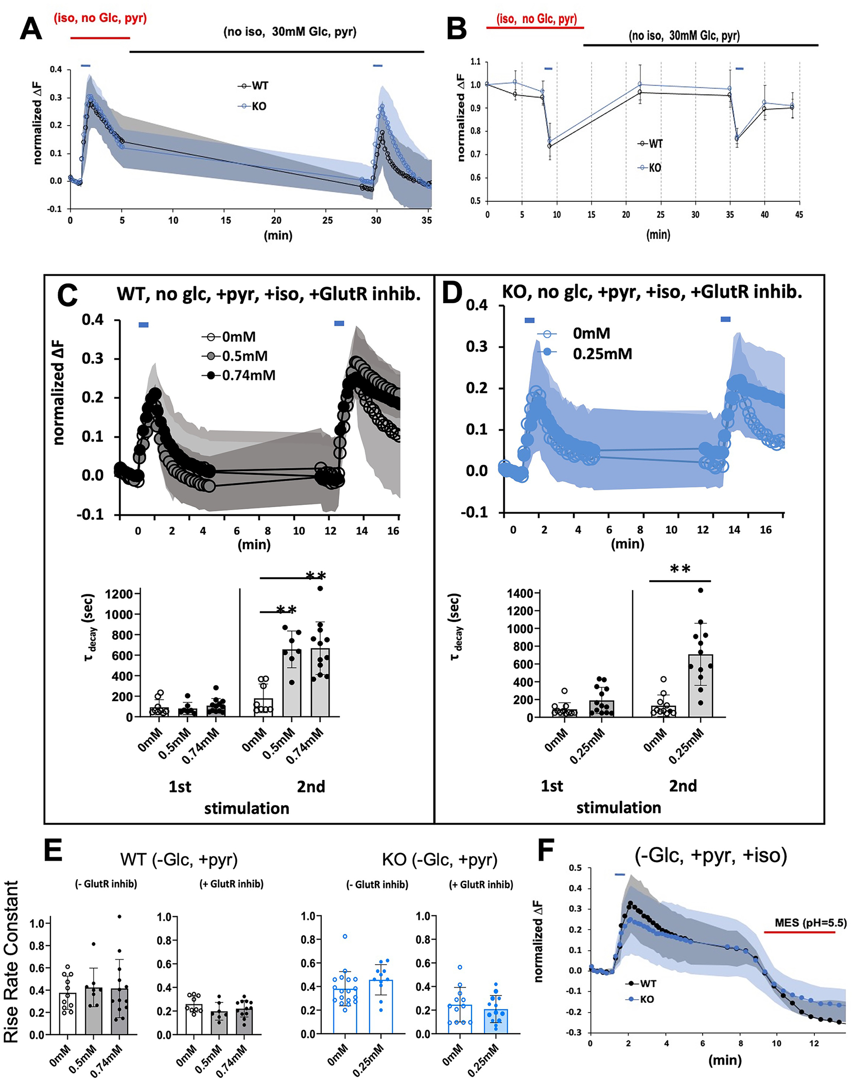
A. Recovery of synaptic recycling following stimulation and removal of isoflurane. Stimulation was as in Figures 1–4. Wildtype and KO cells were exposed to isoflurane at their 2XEC50s in the absence of glucose but with pyruvate (as in Figure 3). Following the first stimulation, isoflurane was removed, and 30mM glucose was added. Endocytosis was re-established following removal of isoflurane and addition of glucose in both genotypes showing reversibility of the anesthetic effects. B. Recovery of ATP levels following stimulation and removal of isoflurane. Stimulation was as in Figures 1–4. Wildtype and KO cells were exposed to isoflurane at their EC95s in the absence of glucose but with pyruvate (as in Figure 3). Following the first stimulation, isoflurane was removed, and 30mM glucose was added. ATP levels recovered to baseline establishing reversibility of the anesthetic effects. C,D. Synaptic Functioning in presence of glutamatergic blockade. To decrease stimulation input, glutamatergic receptors were blocked with antagonists, 6,7-dinitroquinoxaline-2,3-dione (10uM) and 3-(2-Carboxypiperazin-4-yl)propyl-1-phosphonic acid (10uM). C. Wild type cells in 0.5mM or 0.74mM isoflurane in 0mM glucose supplemented with pyruvate. D. Ndufs4(KO) cells in 0.25mM isoflurane in 0mM glucose supplemented with pyruvate. In the presence of isoflurane, endocytosis was markedly defective following the second stimulation. Lower C,D panels. Decay times calculated for WT and KO cells in presence and absence of isoflurane and glutamatergic blockade. Corresponding ATP levels for the WT experiments are shown in Figure S4. E. Histograms showing the effects of isoflurane (WT, 0.5mM and 0.74mM; KO, 0.25mM) on rise time coefficients in the absence or presence of glutamatergic blockade. Left panel represents wildtype cells, right graph represents Ndufs4(KO) cells. F. Stimulating wild type cells in the presence of 0.74mM isoflurane or KO cells in 0.25mM isoflurane in Tyrode buffer (pH 7.4) blocked endocytosis as evidenced by failure of fluorescence to return to baseline. Addition of MES (25 mM, pH 5.5; red bar) to either culture, which acidifies the extracellular compartment, rapidly quenched fluorescence, indicating that pHluorin was exposed to the mild acid.