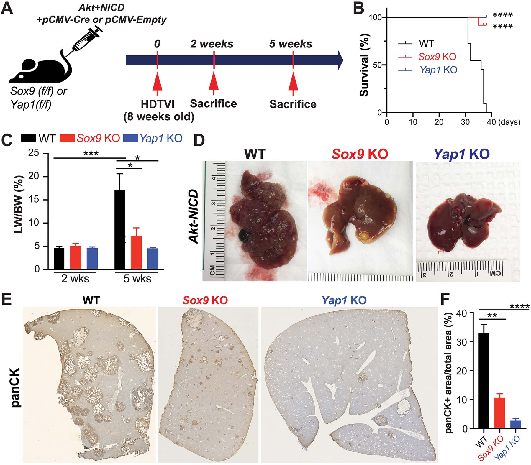
Figure 3. Tumor-specific Sox9 or Yap1 deletion significantly delays Akt-NICD-mediated hepatocyte-derived ICC development.

(A) Experimental design illustrating plasmids used for HDTVI, mice used in study and time-points analyzed. (B) Kaplan–Meier curve showing improved survival of Sox9KO and Yap1KO as compared to WT (C) LW/BW ratio depicts comparable low tumor burden in Akt-NICD Sox9KO, Yap1KO and WT mice at 2w but significantly lower in Sox9KO and Yap1KO at 5w. (D) Representative gross images from Akt-NICD WT show multiple large tumors at 5w, with only occasional small tumor seen in Sox9KO and few gross tumor nodules in Yap1KO at the same time. (E) Representative tiled image of panCK staining in Akt-NICD WT mice, Sox9KO and Yap1KO at 5w and quantification (F). Scale bars:100 μm; error bar: standard error of the mean; *p<0.05; **p<0.01; ***, p<0.001; ****p<0.0001.
