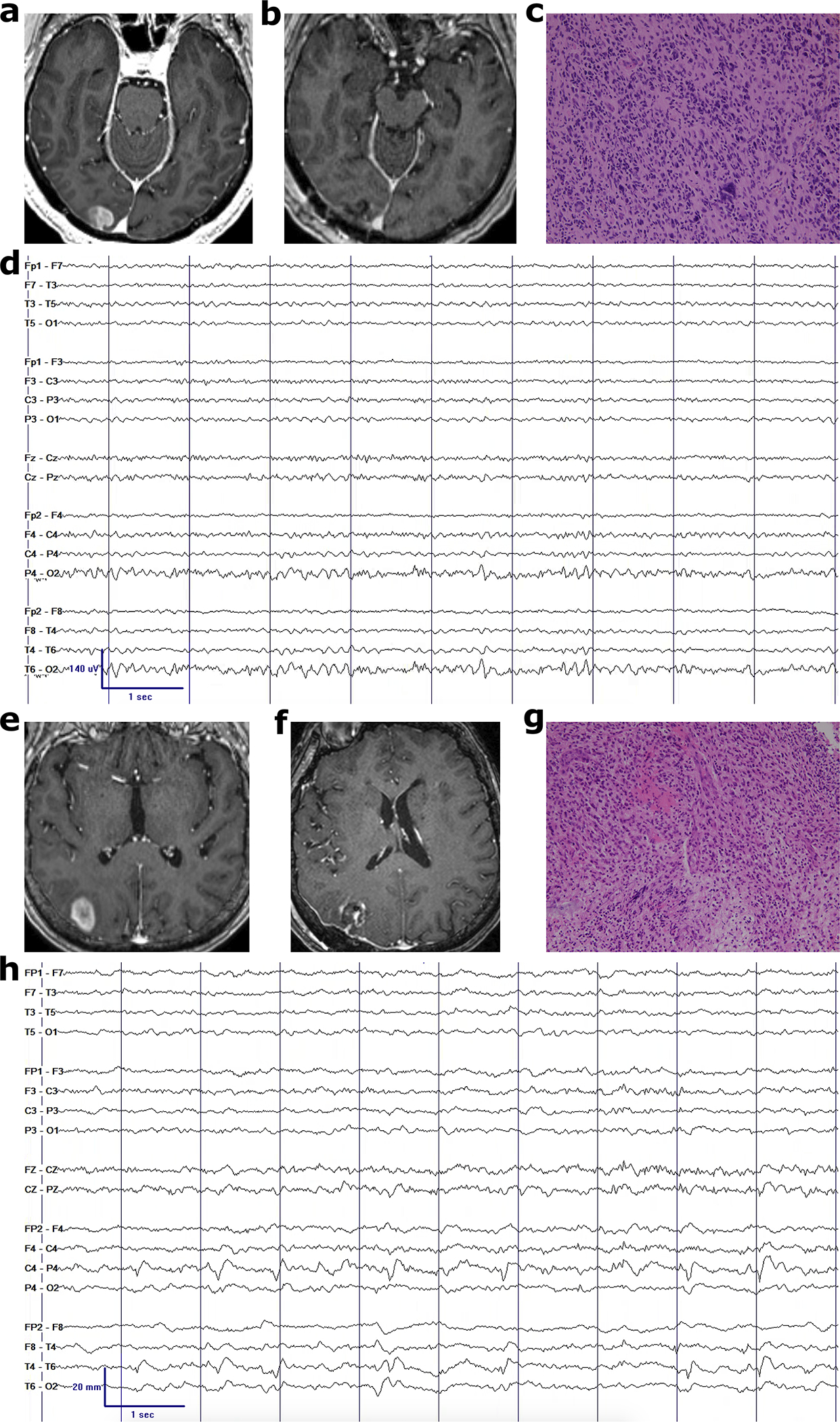
Fig. 2.

Examples of IDH-WT gliomas with and without continuous EEG hyperexcitability. Pre-operative MRI (a,e), post-operative MRI (b,f), representative glioblastoma histology at 20x magnification (c,g), and post-operative EEG (d,h) without and with lateralized periodic discharges in the right posterior peritumoral region, respectively
