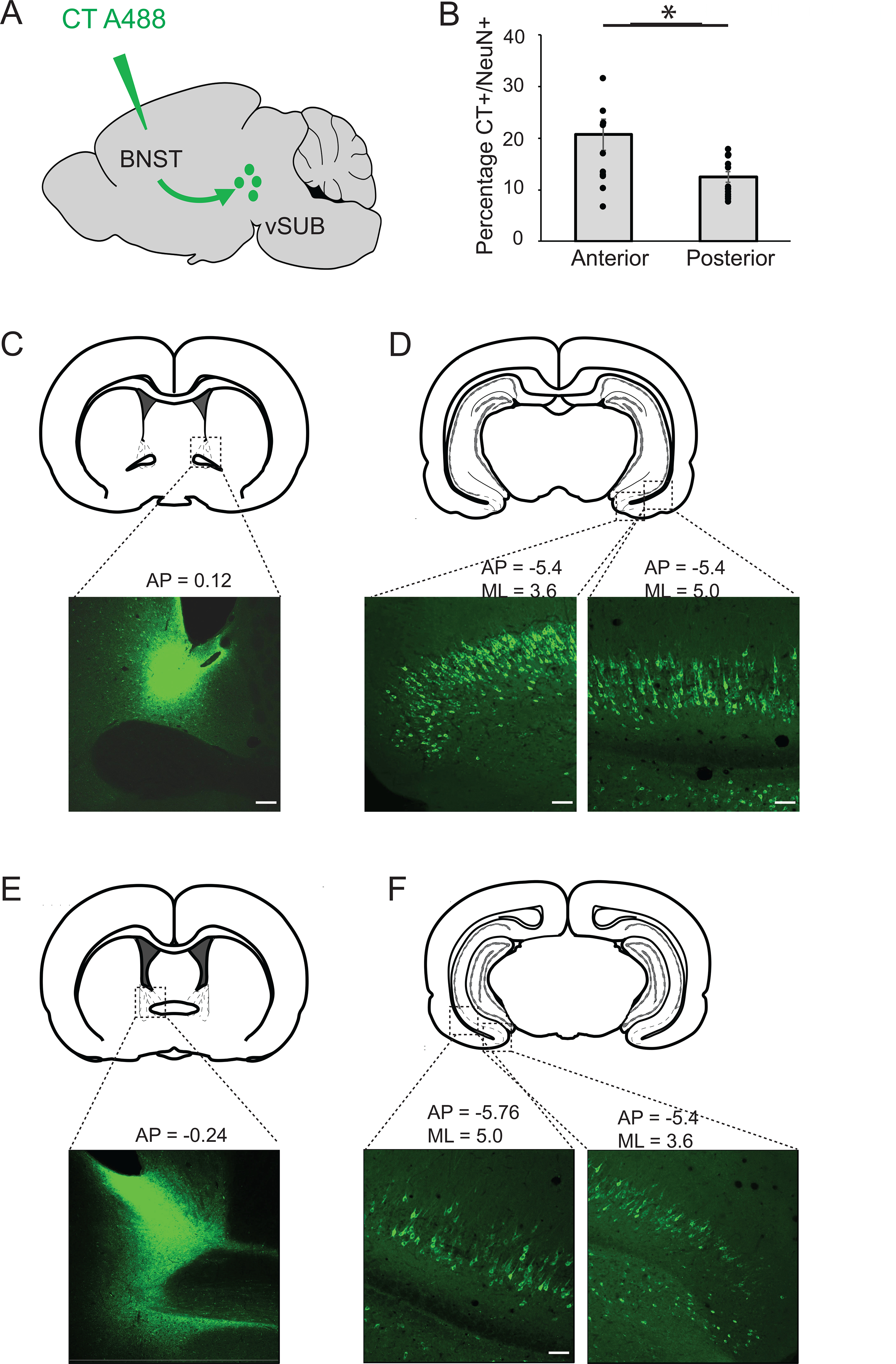
Figure 1:

Topographic organization of projections from the vSUB to the BNST. A: Schematic representation of an injection of cholera toxin (CT) fused to Alexa 488, in the BNST. The toxin is transported retrogradely to label neurons in the vSUB. B: Percentage of anterior versus posterior vSUB neurons projecting to the BNST. Neurons located in the anterior, medial vSUB project to the dorsal anteromedial BNST; neurons located in the posterior, lateral vSUB project to the dorso-postero BNST *p=0.008. C: Example of a CT injection in the dorso antero-medial BNST. D: BNST-projecting neurons located in the anterior, medial vSUB. E: Example of a CT injection in the dorso-posterior BNST. F: BNST-projecting neurons located in the posterior vSUB.
