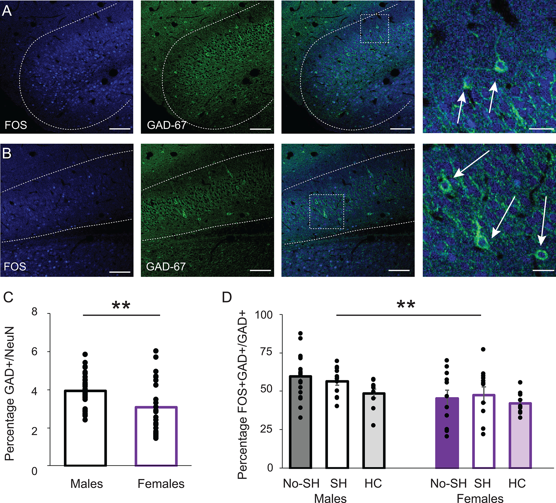
Figure 5:

GABAergic neurons in the vSUB. Confocal images of the anterior medial (A) and lateral (B) vSUB. FOS (left), GAD67 (middle) overlay (right), scale bars =100μm, and magnification (far right) scale bars = 25μm. Arrows point to double-labeled cells. Anterior-posterior distance from Bregma (mm) = −5.5. C: Percentage of GAD+ neurons in the vSUB. Asterisk indicates a significant difference between males and females *p<0.001. B: Double-labeled GAD+/FOS+ cells as a percentage of GAD+ cells in the vSUB in the no shock (NS), shock (S) and home cage (HC) groups in males and females. Asterisk indicates a significant difference between males and females with no difference between behavioral groups *p<0.005.
