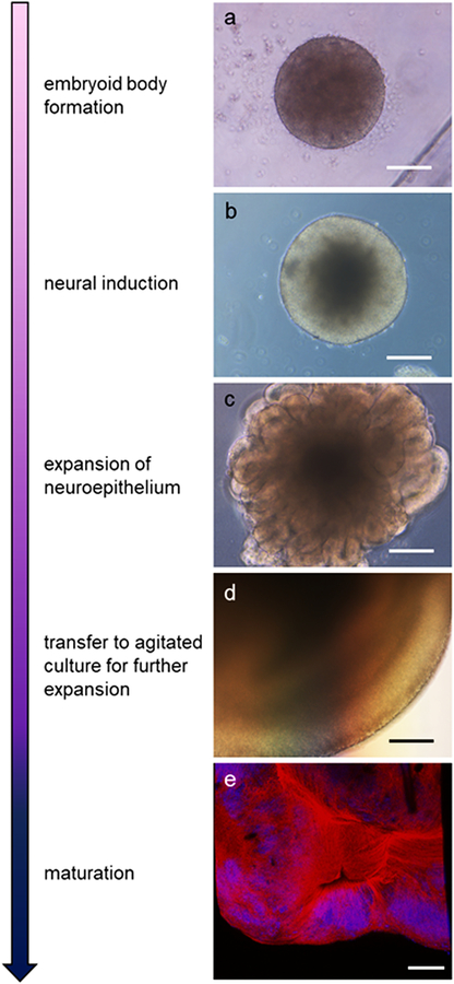
Fig 1.
Organoid developmental stages and morphology. Arrow indicates increasing maturity. Bright-field images show organoid morphology at various developmental stages including; a) fully formed embryoid body beginning neural induction, b) appearance of bright neuroepithelium, c) expansion of neuroepithelial buds, and d) further expansion and structuring of the organoid. e) Detection of mature neurons. Immunofluorescent staining shows neurofilament light chain (red) and nuclei (blue; DAPI) at approximately 45 days old. Scale bar = 100 μm

