Figure 1.
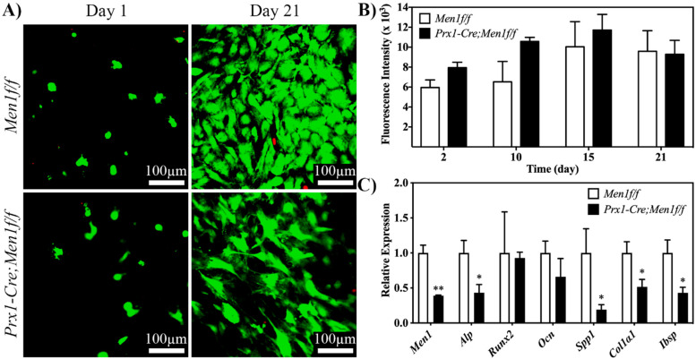
Viability, proliferation and osteogenic differentiation of calvarial osteoblasts from 6 month old Men1f/f and Prx1-Cre, Men1f/f mice seeded in 3D dense collagen gels under osteogenic medium in vitro. (A) Confocal images of calcein-AM and EthD-1-stained cells at days 1 and 21. No qualitative differences between the two groups were observed (n = 3). (B) Metabolic activity of seeded cells was assessed at days 2, 10, 15 and 21 of osteogenic differentiation. Seeded gels were stained in osteogenic medium with 10% alamarBlue® reagent, and fluorescence was determined using a microplate reader. No significant differences between the two groups were observed (n = 3). (C) Expressions of Men1, Alp, Runx2, Ocn, Spp1, Col1α1 and Ibsp, of seeded cells at day 21. Significantly (* p < 0.05 and ** p < 0.01) lower expressions of markers were observed in Prx1-Cre, Men1f/f compared to Men1f/f osteoblasts (n = 3).

