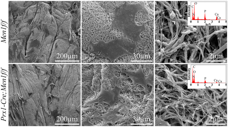
Figure 2.
SEM micrographs at day 21 of calvarial osteoblasts from 6 month old Men1f/f and Prx1-Cre, Men1f/f mice 3D seeded in 3D dense collagen gels under osteogenic medium in vitro. Lower magnification SEM micrographs (left panel) depicted evenly distributed cells within the collagen gels in both groups. Higher magnification images qualitatively indicated that the morphology of Men1f/f cells appeared larger compared to that of Prx1-Cre, Men1f/f cells (middle panel). While mineralized particles were detected in both groups, mineralized collagen fibrils were only indicated in Men1f/f seeded gels (right panel). EDS revealed the presence of calcium and phosphorous in both groups (inset in right panel) (n = 3).

