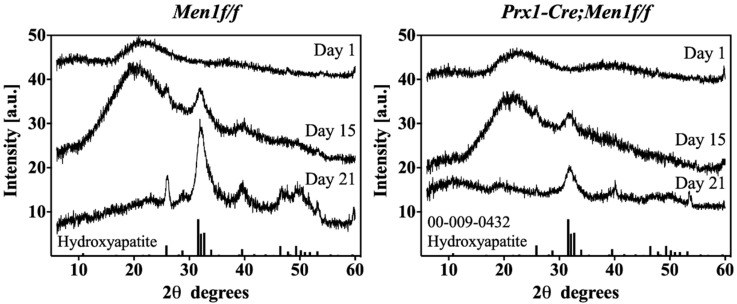
Figure 4.
XRD diffractograms of cell-mediated matrix mineralization. Left and right diffractograms show time-dependent XRD diffractograms of 3D seeded calvarial osteoblasts from 6 month old Men1f/f and Prx1-Cre, Men1f/f, respectively, seeded in dense collagen gels up to day 21 in osteogenic differentiation medium. Both groups displayed a time-dependent amorphous-to-crystalline transition. Men1f/f seeded gels displayed higher intensity peaks relative to those of Prx-Cre, Men1f/f (n = 3).

