Figure 3.
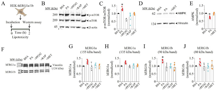
Western blot assay revealed modulation of mTOR, AMPK, and hERG1a/1b expression in lipotoxic HEK293 cells. (A) Experimental protocol: mTOR, AMPK, and hERG1a/1b expression in HEK293 cells under control conditions and after pre-exposure to lipotoxicity for 2 h was measured. (B) Western blot assays show the levels of activated mTOR (p-mTOR) induced by lipotoxicity in the absence or presence of mTOR inhibitors (PP242 and RAP), or AMPK activator (MET), compared to in control cells. (C) Quantification of the Western blot and statistical analysis showing the effects of PP242, RAP, and MET on the levels of p-mTOR. (D,E) Western blot and quantification/statistical analysis of AMPK expression in control and lipotoxic cells in the presence or absence of MET. (F–J) Western blot and quantification/statistical analysis showing the levels of hERG1a and 1b expression in the absence and presence of PP242, RAP, and MET in control and lipotoxic cells. (* statistical significance at p < 0.05).

