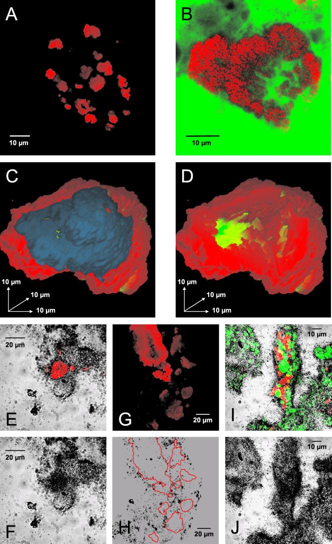
FIG. 4.
In situ analyses of Nitrospira-like bacteria within activated sludge and biofilms. (A) Nitrospira cell aggregates detected in activated sludge by FISH with probe S-G-Ntspa-0662-a-A-18 (red). (B) Nitrospira cell aggregate detected in biofilm from reactor SBBR 1 by FISH with probe S-G-Ntspa-0662-a-A-18 (red). All of the Nitrospira colonies detected in SBBR 1 with probe S-G-Ntspa-0662-a-A-18 also hybridized with probe S-*-Ntspa-0712-a-A-21 (data not shown). The biofilm was also stained with fluorescein (green). (C and D) Three-dimensional reconstruction of a Nitrospira cell aggregate from reactor SBBR 1 stained by FISH with probe S-G-Ntspa-0662-a-A-18. Nitrospira cells are red; the blue part of the microcolony in panel C was digitally removed to allow insight into the aggregate (D). Voids within the aggregate are green. (E and F) Uptake of bicarbonate by Nitrospira-like bacteria in biofilm from reactor SBBR 1 under aerobic incubation conditions. (E) Nitrospira cells stained by probe S-G-Ntspa-0662-a-A-18 (red) combined with the micrograph of the radiographic film at the same position. Panel F shows only the film to visualize the MAR signal at the position of the Nitrospira cells. Other MAR signals were caused by CO2-fixing bacteria, which were not detected by the Nitrospira-specific probe. (G and H) No uptake of acetate by Nitrospira-like bacteria in biofilm from reactor SBBR 1 under aerobic incubation conditions. (G) Nitrospira cells stained by probe S-G-Ntspa-0662-a-A-18 (red). Panel H is a micrograph of the radiographic film at the same position. The localization of the Nitrospira microcolonies in panel G is indicated by red borderlines in panel H. (I and J) Uptake of pyruvate by Nitrospira-like bacteria (stained by probe S-G-Ntspa-0662-a-A-18; red) and by ammonia oxidizers (stained by probes NEU and Nso1225; green) in biofilm from reactor SBBR 1 under aerobic incubation conditions. The fluorescence recorded in a stack of images by the CLSM is combined by orthographic projection in panel I. Stacked cells of Nitrospira-like bacteria and ammonia oxidizers appear therefore yellow. The image stack was acquired to ensure that all of the nitrifiers that contributed to the MAR signal would be visible in the final image.

