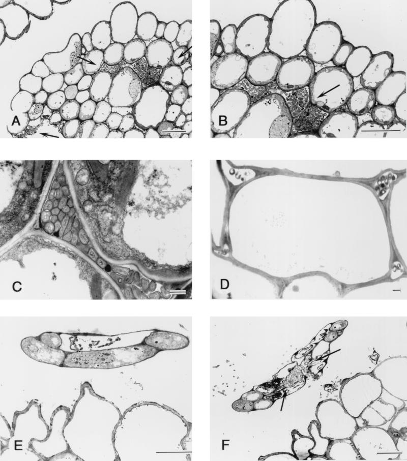
FIG. 3.
Transmission electron micrograph of transverse section through rice plant of O. officinalis W0012 seven days after seed inoculation with Herbaspirillum sp. strain B501gfp1. (A) The bacteria have invaded the shoot tissue which is a unexpanded third leaf (indicated by the two top arrows), and some bacteria were outside the leaf tissue (the lowest arrow). Bar = 10 μm. (B) High-magnification view of panel A showing bacteria colonizing the intercellular space within the rice tissue. Bar = 10 μm. (C) The bacteria colonizing the intercellular space of the third leaf of rice. Bar = 1 μm. (D) Section from rice coleoptile, showing the bacteria colonizing the intercellular space but not densely populated. Bar = 1 μm. (E) Cross section from the tip of a fourth leaf of rice shoot, showing little invasion of the undifferentiated leaf, which has only five cells. Bar = 10 μm. (F) A lower cross section from the same fourth leaf tip seen in panel E. The bacteria have entered the young fourth leaf and colonized the intercellular space (arrow). Bar = 10 μm.

