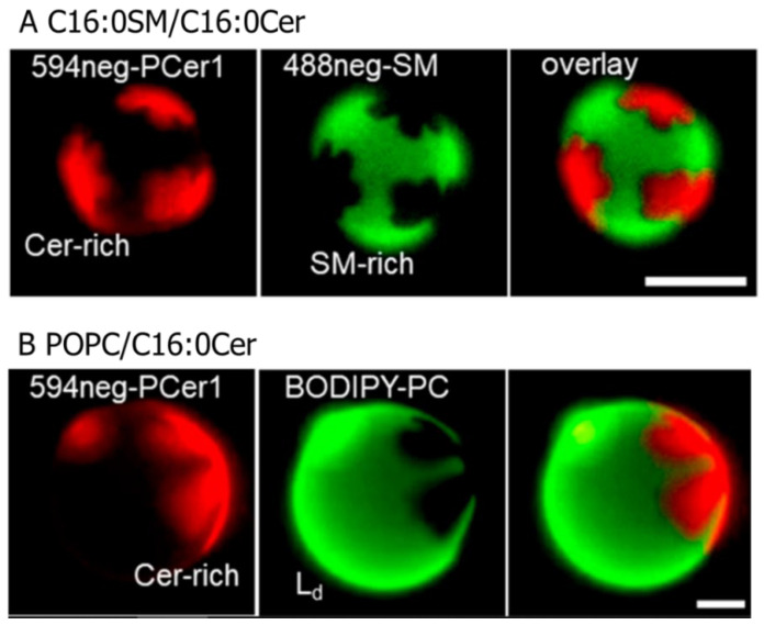
Figure 2.
Fluorescence micrographs of binary-component giant unilamellar vesicles (GUVs) that underwent phase separation between the ceramide-rich and ceramide-poor (thus, phospholipid-rich) domains. (A) C16:0SM/C16:0Cer (95:5, molar ratio) GUVs containing 0.2 mol % 594neg-PCer1 (ceramide-rich domain marker) and 0.2 mol % 488neg-SM (SM-rich domain marker). (B) Palmitoyl-oleoyl–phosphatidycholine (POPC)/C16:0Cer (95:5, mole ratio) GUVs containing 0.2 mol % 594neg-PCer1 and BODIPY-PC (POPC-rich fluid phase marker). Bars indicate 10 μm. Image brightness and contrast were adjusted for clarity. This figure was adapted with permission from [41]. Copyright 2019 American Chemical Society.

