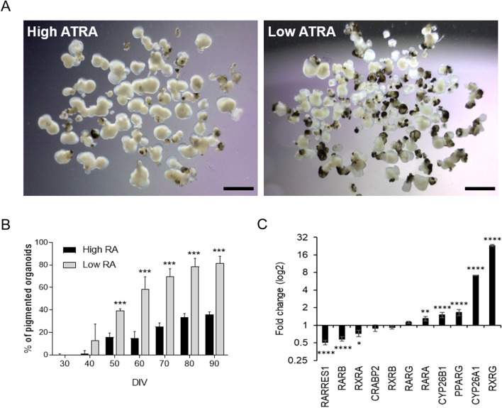
Fig. 1.
All-trans retinoic acid modulates pigmentation of hiPSC-derived multiocular organoids. A Representative images of multiocular organoids cultured in suspension in low or high all-trans retinoic acid (ATRA) concentrations at day 90. Scale bars: 3 mm. B Quantification of the number of pigmented ocular organoids at several days in vitro (DIV) is shown as mean ± SD (starting with n = 420 organoids (high ATRA) and n = 512 organoids (low ATRA) divided in 6 replicates). Asterisks represent statistical significance from a Student’s t test (***p < 0.0001). C Quantitative PCR analysis of ATRA signaling pathway components in multiocular organoids. Values are normalized to GAPDH and expressed as 2−ddCt (log2 scale). Data presented as mean ± SD (n = 6–10 organoids, three replicates). Values indicated by stars are significantly different from those in low ATRA (two-way ANOVA with Sidak’s multiple comparison test; *p < 0.05; **p < 0.001; ****p < 0.0001)

