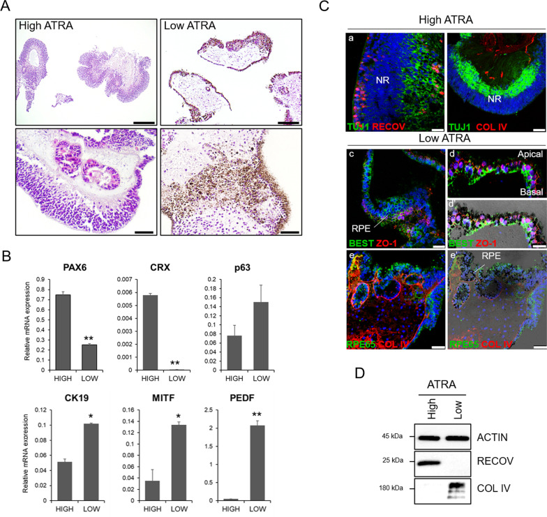
Fig. 2.
High ATRA promotes neuroretinal organoids, while low ATRA induces pigmented epithelial organoids. A Hematoxylin and eosin staining of paraffin sections from the multiocular organoids showing predominant neuroretinal (NR) organoids in high ATRA condition and pigment epithelial (PE) organoids in low ATRA condition at day 90. Scale bars: 250 µm (top images); 50 µm (bottom images). B Gene transcripts analysis by quantitative PCR in multiocular organoids. Values are normalized to GAPDH. Data presented as mean ± SD (n = 6–10 organoids, 3 replicates). Values indicated with stars are significantly different from those in high ATRA conditions (Student’s t test; *p < 0.01; **p < 0.001). C Confocal (a–e) and bright-field merged (e’) images of multiocular organoid paraffin sections stained with neuroretinal markers recoverin (RECOV) and TUJ1, and RPE markers bestrophin 1 (BEST), zonula occludens 1 (ZO1) and RPE65, and stromal collagen type IV (COL IV)). Nuclei are stained with DAPI. Scale bars: 50 µm in b,e,e’; 25 µm in a,c; 10 µm in d,d’. D Western blot analysis of RECOV and COL IV protein expression. Actin was used as the loading control

