Fig. 4.
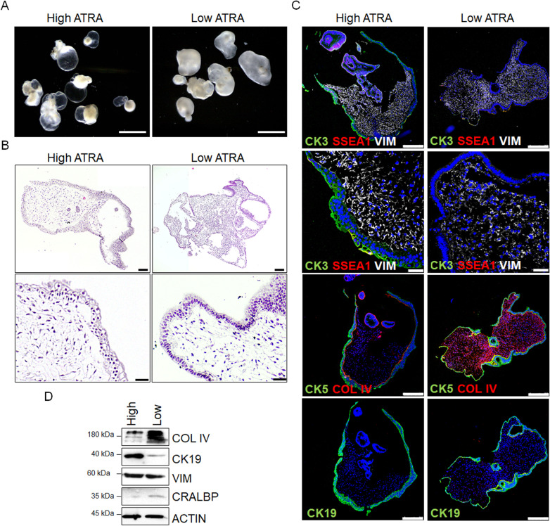
High ATRA concentration increases corneal organoids transparency and decreases collagen deposition in the stroma. A Representative images of corneal organoids at day 120 cultured in high ATRA (transparent corneal organoids) and low ATRA (opaque corneal organoids) concentrations. Scale bars: 3 mm. B Hematoxylin and eosin staining of paraffin sections of transparent and opaque corneal organoids. Scale bars: 150 µm (upper panels); 50 µm (lower panels). C Immunofluorescent images of transparent (high ATRA) and opaque (low ATRA) corneal organoids expressing corneal markers CK3, corneal-conjunctival markers CK5 and MUC1, conjunctival marker CK19, and limbal stem cell marker SSEA1. Stromal cells are stained with vimentin (VIM) and collagen type IV (COL IV). Nuclei are stained with DAPI. Scale bars: 50 µm. D Western blot analysis of protein levels related to stroma (COL IV and VIM), cornea-conjunctiva (CK19), and RPE (cellular retinaldehyde–binding protein, CRALBP) in corneal organoids. Actin was used as a loading control

