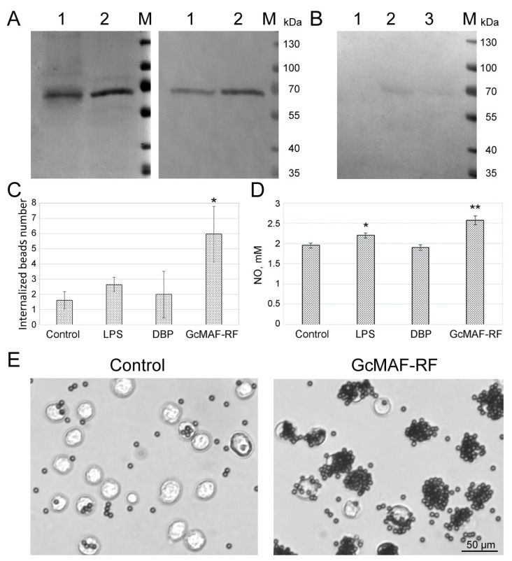
Figure 1.
Characterization of DBP and DBP-derived GcMAF-RF. (A) Polyacrylamide gel of DBP samples obtained using actin-agarose (1), and Sepharose (with 25-hydroxyvitamin D3) (2) affinity chromatography (left) and Western blot of these samples with antibodies against Gc group proteins (right). (B) Western blot of a polyacrylamide gel for Helix pomatia lectin. 1—DBP, 2—GcMAF-RF, and 3—FBS. M—Thermo ScientificTM Page RulerTM Prestained protein Ladder molecular marker (Thermo Fisher Scientific Inc., Waltham, MA, USA). (C) Phagocytic activity of murine PMs treated with LPS, DBP, and GcMAF-RF compared to control untreated macrophages. Data are presented as Mean ± SD (n = 4), * differences compared to the control are significant, p < 0.05 (Mann–Whitney U test). (D) NO production by murine PMs treated with LPS, DBP, and GcMAF-RF compared to control untreated macrophages. Data are presented as Mean ± SD (n = 5), differences compared to the control are significant, * p < 0.05, ** p < 0.01 (Mann–Whitney U test). (E) Images of the beads phagocytized by naïve macrophages (control) and macrophages exposed to GcMAF-RF.

