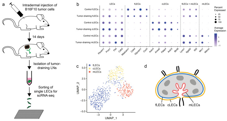
Figure 1.
Single-cell RNA sequencing (scRNA-seq) shows persistence of lymphatic endothelial cell (LEC) subsets in lymph nodes (LNs) draining tumors. (a) Experimental workflow. B16F10 melanoma cells were injected intradermally into the flanks of C57Bl/6N mice. After 14 days, mice were sacrificed and tumor-draining inguinal LNs collected. LNs were digested enzymatically, the resulting single cell suspension stained for lymphatic markers, and individual LECs sorted by FACS into 384-well plates for scRNA-seq. (b) Dot plot showing expression of established LEC and subset marker genes in subcapsular sinus floor LECs (fLECs), subcapsular sinus ceiling LECs (cLECs) and medullary LECs (mLECs) from tumor-draining and control LNs. (c) UMAP visualization of all 581 LN LECs used for analysis, 225 from control and 356 from tumor-bearing mice. Cells are colored by cluster; fLECs (blue), cLECs (yellow), and mLECs (red). (d) Schematic illustration of LEC subtypes within the LN.

