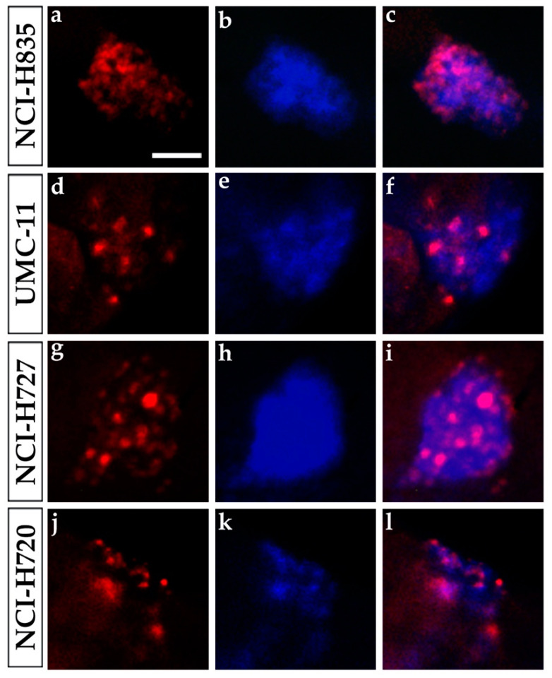
Figure 5.
Ki-67 immunostaining of lung carcinoid grafted embryos. Representative images of 48 hpi Tg(fli1a:EGFP)y1 zebrafish embryos at the level of the graft regions after immunofluorescence assay to detect Ki-67 localization (red). Embryos were implanted with NCI-H835 (a–c), UMC-11 (d–f), NCI-H727 (g–i), and NCI-H720 (j–l) cells. Injected lung carcinoid cells were previously blue-stained (b,e,h,k). The merge of red and blue channels (c,f,i,l) showed that Ki-67 staining is tumor cell specific. Scale bar: 50 µm.

