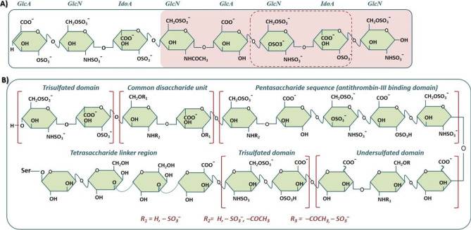
Fig. 1.
A Heparin with a representative polysaccharide containing four disaccharide building blocks composed of one uronic acid (UA) and one glucosamine (GlcN) moiety. The disaccharide sequence — GlcNS3S6S-IdoA2S — in the dashed frame (FG) constitutes the highly sulfated region and major repeating structural unit within heparin while the block-shaded pink is the pentasaccharide sequence (or antithrombin II–binding domain). One of the two UA residues (iduronic acid, IdoA) present in the pentasaccharide sequence is consistently sulfated at the C-2 position, whereas the hydroxyl groups (OH) at both C-2 and C-3 of the other uronic moiety (glucuronic acid, GlcA) are unsubstituted. B A representative heparin comprising (left to right continuing to lower panel): a trisulfated domain, a common disaccharide unit, a pentasaccharide sequence (antithrombin III–binding domain), a trisulfated domain, and a tetrasaccharide linker region, GlcA-Gal-Gal-Xyl-Ser (source: Wang and Chi [5] Recent advances in mass spectrometry analysis of low molecular weight heparins. J. Chin. Chem. Lett., 2018, 29(1): 11–18; Ekre et al. Use of chemically modified heparin derivatives in sickle cell disease. US9480702 (2016))

