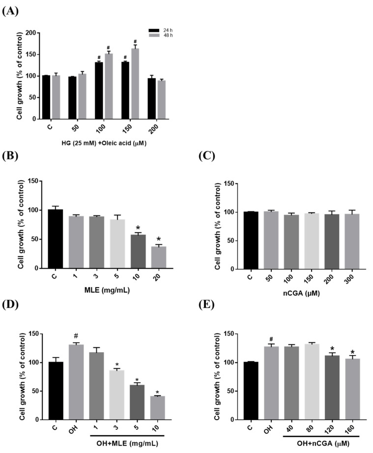
Figure 1.
Effect of mulberry leaf extract (MLE) and neochlorogenic acid (nCGA) treatment on cell viability/proliferation. (A) A7r5 cell proliferation after being induced by HG (25 mM) and Oleic acid (0.5, 1.0, 1.5 and 2.0 mM) for 24 and 48 h. (B,C) A7r5 cells were treated with indicated concentrations of MLE (1.0, 3.0, 5.0, 10, and 20 mg/mL) and nCGA (0, 50, 100, 150, 200 and 300 μM) for 24 h. (D,E) A7r5 cells were pretreated with OH and then exposed to different concentrations of MLE (0.0, 1.0, 3.0, 5.0, and 10 mg/mL) and nCGA (0, 40, 80, 120, and 160 μM) for 24 h. Cell viability/proliferation of the A7r5 cells was evaluated using the 3-(4,5-dimethylthiazol-2-yl)-2,5 diphenyltetrazolium bromide (MTT) assay. The quantitative data are presented as the mean ± SD from a minimum of three independent experiments. # p < 0.05 as compared to the C (control). * p < 0.05 as compared to OH group.

