Figure 3.
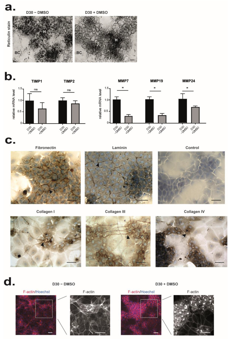
Effects of DMSO on matrix remodeling and cell organization. (a) ECM deposition was detected by reticulin staining on HepaRG cells differentiated from D15 of culture in the presence (D30+ DMSO) or the absence of DMSO (D30 − DMSO). (b) Microarray data for TIMP1, TIMP2, MMP7, MMP19, and MMP24 are expressed relative to conditions D30 − DMSO, arbitrary set to 1. The significance of DMSO treatment (D30 + DMSO vs D30 − DMSO) was evaluated by an unpaired t-test * p < 0.05, ns: not significant. (c) Immunostaining of fibronectin, laminin, collagen type I, III, and IV in HepaRG cells differentiated in the presence of DMSO (D30+ DMSO). Immunolocalization without primary antibody was performed as control. (d) Immunolocalization of F-actin in HepaRG cells differentiated from D15 of culture in the presence (D30+ DMSO, left panel) or in the absence of DMSO (D30 − DMSO, right panel). Nuclei were visualized by staining with Hoechst (shown in blue). Scale bars = 7 µm.

