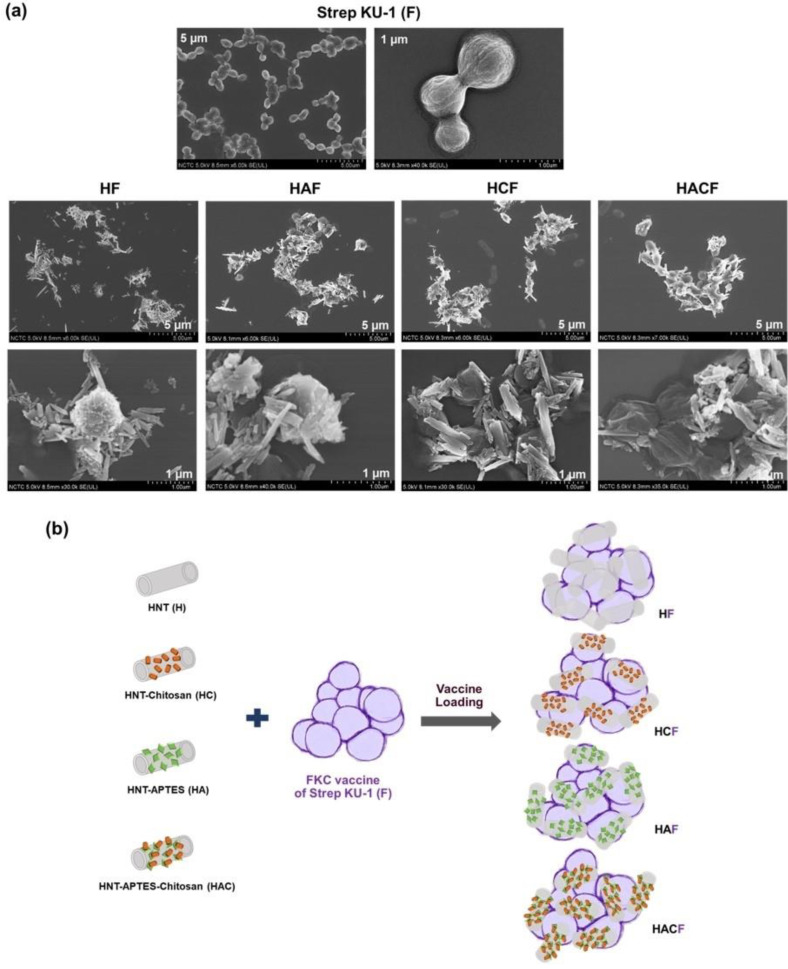
Figure 1.
Scanning Electron Microscopy (SEM) observation and schematic diagram of Strep KU-1 and HNTs: (a) SEM images of the bared Strep KU-1 molecules (F) as well as the complexes of Strep KU-1 combined with HNTs (HF, HAF, HCF and HACF) at scale bars of 1 µm and 5 µm. (b) The schematic diagram proposing the surface binding characteristics of HNT moiety on the Strep KU-1 surface.

