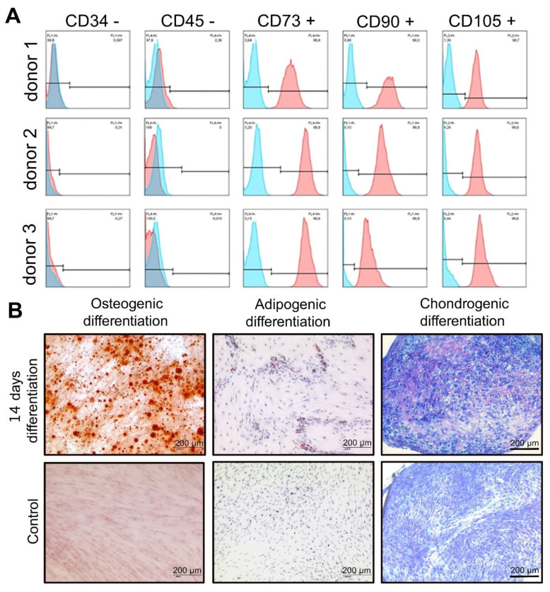
Figure 1.
Characterization of primary human PDL cells. (A) hPDL cells were first characterized by flow cytometry analysis of cell surface markers. The expression pattern for all donors is CD34−, CD45−, CD73+, CD90+ and CD105+ (blue color: isotype control, red color: surface marker) and (B) hPDLs differentiation towards adipocytes, chondrocytes and osteoblasts was performed. hPDL cells were grown in osteogenic, adipogenic and chondrogenic differentiation media for 14 days and then stained with alizarin red, Oil red O and Toluidin blue, respectively. The red color represented mineralized calcium depositions after PDL cells differentiated into osteoblast-like cells. A large number of orange lipid vacuoles were seen in PDL cells after culture in AIM and staining with Oil red O. Staining with Toluidine blue revealed excessive production of extracellular matrix components and proteoglycans during the culture process with CIM (scale: 200 μm).

