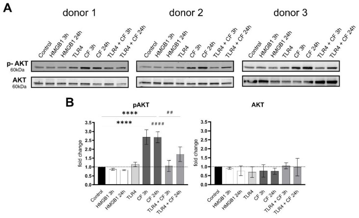
Figure 4.
Compressive forces upregulate phosphor AKT in primary hPDL cells. (A) Protein production levels and phospho-AKT were determined by Western blot in different conditions: HMGB1 (100 ng/mL), TLR4 blocking antibody (5 µg/mL) (TLR4: TLR4 blocking antibody) and compressive force (CF) 2 g/cm2 at 3 and 24 h. Three different donors showed similar patterns. Under all conditions, AKT showed no differences, but the phosphorylated AKT was significantly upregulated with compressive forces (CF) for 3 h and 24 h. Compressive forces with additional blocking TLR4 monoclonal antibody led to significant downregulation comparable to the control. (B) Quantification of three donors, normalized to the control with stain-free technology. CF conditions without TLR4 blocking antibody showed a significant upregulation. Data were tested for normal distribution by Shapiro–Wilk test. Afterward, a one-way analysis of variance (ANOVA) followed by Tukeys’ post hoc test was performed. Statistically significant differences to control are marked by asterisks (**** p < 0.0001) and hashtag show significant differences between CF and TLR4 +CF (## p < 0.01; #### p < 0.0001).

