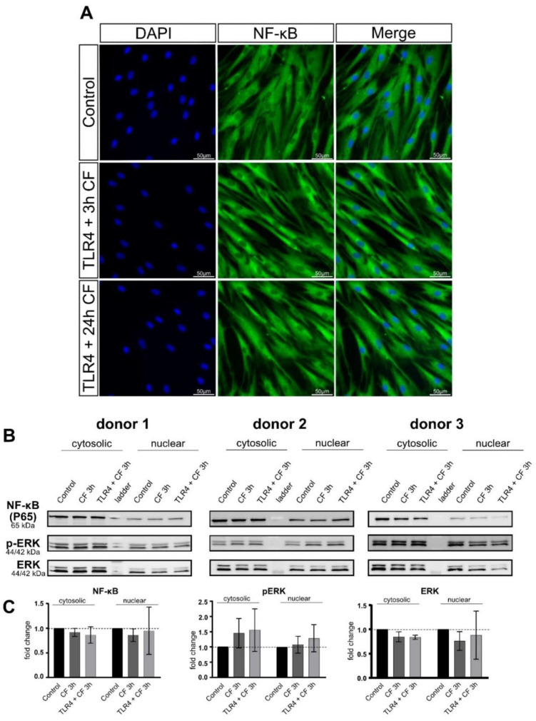
Figure 6.
Fluorescence images of CF and TLR4 blocking antibody on PDL cells. (A) PDL cells treaded with and without TLR4 blocking antibody (5 µg/mL) and compressive force 2 g/cm2 for 24 h. NF-kB was stained green (Alexa 488), blue areas represent nuclei (DAPI); scalebar 50 µm, CF: compressive force NF-kB was not translocated to the nucleus in any condition. (B) Protein production of NF-kB, ERK and phospho-ERK were determined by Western blot. (C) Quantification of three donors, normalized to the control with stain-free technology. CF conditions without TLR4 antibody showed a significant upregulation for phospho-ERK and phospho-p38.

