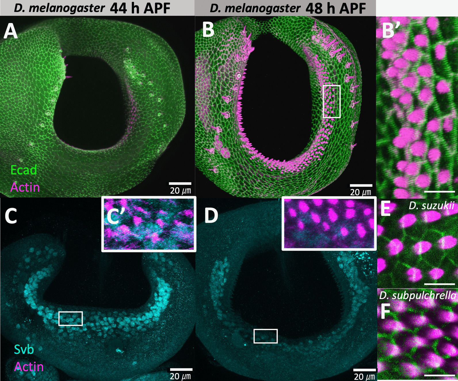
Fig. 3.

Confocal images of developing ovipositors in D. melanogaster (y, w), D. suzukii (TMUS08), and D. subpulchrella (H243). A Configuration of E-cadherin (green) and F-actin/phalloidin (magenta) at 44 h APF in D. melanogaster. B Configuration of E-cadherin (green) and F-actin/phalloidin (magenta) at 48 h APF in D. melanogaster. B’ Enlarged image of the rectangular area in (B). C Immunostaining of Svb (light blue) at 44 h APF in D. melanogaster. C’ Enlarged image of the rectangular area in (C) with F-actin/phalloidin (magenta). D Immunostaining of Svb (light blue) at 48 h APF in D. melanogaster. D’ Enlarged image of the rectangular area in D with F-actin/phalloidin (magenta). E Configuration of E-cadherin (green) and F-actin/phalloidin (magenta) in D. suzukii at 45 h APF. F Configuration of E-cadherin (green) and F-actin/phalloidin (magenta) in D. subpulchrella at 45 h APF. Scale bars indicate 20 μm in (A–D) and 5 μm in (B’), (E), and (F)
