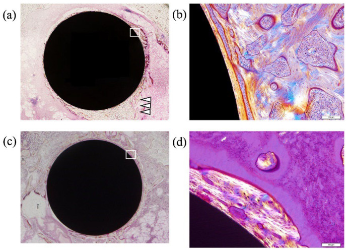
Figure 10.
Non-decalcified histological assessment of TiNbSn implanted femurs. TiNbSn rods anodized with sulfuric acid, and untreated TiNbSn rods were implanted in rabbit femurs. Histological assessment was performed six weeks after TiNbSn rod implantation. Arrowheads indicate new bone formation. (a) Low-magnification image of anodized TiNbSn rod groups. (b) Low-magnification image of untreated TiNbSn rod group. (c) High-magnification image of anodized TiNbSn rod group. (d) High-magnification image of untreated TiNbSn rod group.

