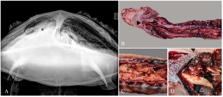
Figure 1.
Necrotizing tracheitis and bronchopneumonia due to Serratia proteamaculans infection. Imaging and gross pathology. (A) Extensive areas of increased soft tissue opacity (*) were present bilaterally on craniocaudal radiographs. (B) From the glottis (►), the tracheal mucosa was extensively ulcerated (∇) and replaced by diphtheritic membranes composed of fibrin and necrotic debris. (C) Ulcerated tracheal mucosa filled with diphtheritic membrane (*), and (D) Bronchi partially occluded with a diphtheritic membrane plug (Δ) and hemorrhagic pulmonary parenchyma (*).

