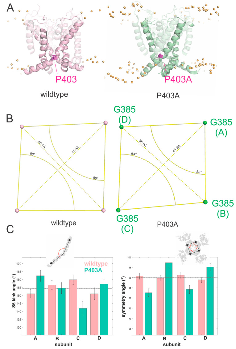
Figure 5.
(A) Side view of the homology model of the Kv1.1 channel built upon the crystal structure of Kv1.2/2.1 chimera showing the localization of the P403 residue and P403A mutation. P403A is highlighted in magenta. (B) Loss of symmetry of wildtype and P403A measured at the top of S6 (position G385). (C) Measure of the S6 kink angle (left) and symmetry angle (right) for the four subunits of WT and P403A channels. The kink angle of the S6 was measured as the angle between the helix axis of the S6 above and below the PVP/A-motif. The symmetry angles are formed by the vectors from G385 of one subunit to the G385 of the two neighboring subunits. Values are given as mean ± SD of the values measured every 0.1 ns over the last 50 ns.

