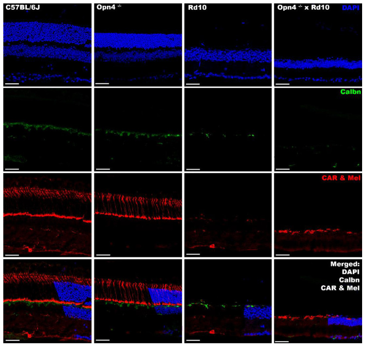
Figure 7.
Immunohistochemical labeling of cones and ipRGCs in different models. From left to right, the different animal models are presented (C57BL/6J; Opn4−/−; Rd10; Opn4−/− × Rd10). From top to bottom, the different agents and antibodies used are presented. DAPI: intercalating marker of cell nuclei; Calbindin (Calbn): protein located in the horizontal cells; Cone Arrestin (CAR): cone labeler; Melanopsin (Mel): melanopsin labeler, located in the ipRGCs; ipRGCs are marked by white arrowhead. The bottom row shows: “Merged” of different labelings. Opn4−/− × Rd10 animal model showed total absence of cells with photosensitive capacities. The scale bar indicates 50 µm.

