Figure 1.
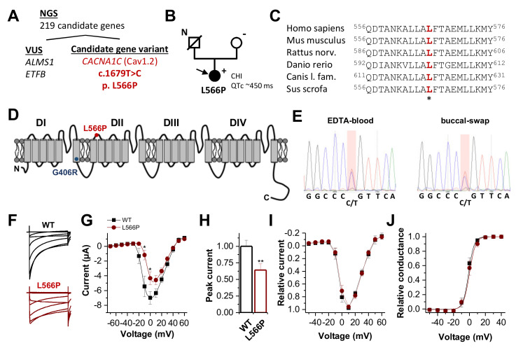
CaV1.2 channel topology, localization and conservation of the CaV1.2L566P mutation identified by WES and electrophysiological characterization. (A) NGS panel sequencing of 219 genes associated with familial hyperinsulinism or related disorders of the glucose metabolism variants filtered out four variants of uncertain significance (VUS) and one candidate gene variant in CACNA1C. (B) Pedigree of the family. N, no genomic data available; +, heterozygous carrier of c.1679T > C; −, absence of the variant. The filled circle and arrow indicate the diseased index patient. CHI, congenital hyperinsulinism. (C) Partial amino acid sequence alignment of CaV1.2, illustrating the conservation of the respective leucine residues among the different orthologues. (D) CaV1.2 channel topology. DI to DIV indicate the four calcium channel domains, each consisting of six transmembrane segments and one pore-forming domain. The localization of the channel variants CaV1.2L566P and CaV1.2G406R is indicated as red and blue dots, respectively. (E) Electropherogram of the mutation carrier confirming the CACNA1C variant by Sanger sequencing of DNA isolated from EDTA blood and buccal swabs of the patient. (F) Representative current traces of CaV1.2WT (black) and CaV1.2L566P (red) recorded from a holding potential of −80 mV, with voltage steps of 1 s duration ranging from −70 to +60 mV in 10 mV increments. The interval between the steps was 30 s. (G) Current–voltage relationship of wild-type CaV1.2 (black) (n = 19) and CaV1.2L566P (red) (n = 14) channels recorded from three independent batches of oocytes. (H) Analysis of the relative peak current amplitudes normalized to wild-type CaV1.2. (I) Relative current–voltage relationships of wild-type CaV1.2 (black) (n = 10) and CaV1.2L566P (red) (n = 10) normalized to the peak current. In this analysis, only recordings with peak current amplitudes <5 µA were considered. (J) Conductance–voltage relationship of CaV1.2WT and CaV1.2L566P (red). **, p < 0.01; *, p < 0.05.

