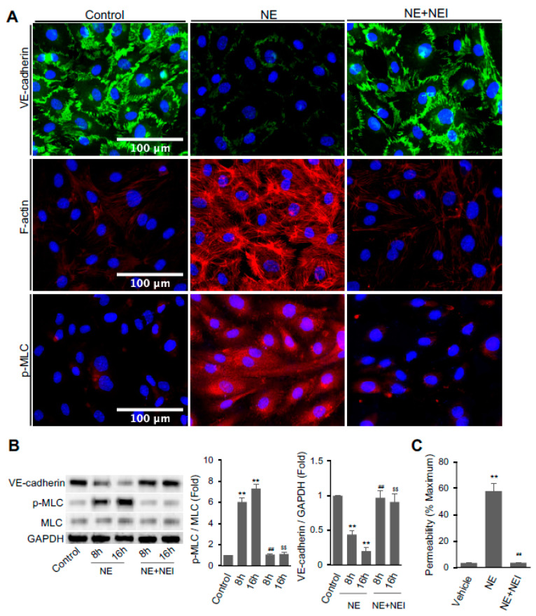
Figure 1.
Neutrophil elastase (NE) decreased VE-cadherin protein and increased F-actin formation, MLC phosphorylation, and permeability in hECs. (A) Confluent hECs were treated with the vehicle or 20 nM NE in the presence or absence of NE inhibitor GW311616A (NEI, 1 μM) for 16 h. Cells were fixed and fluorescence labeled with antibodies against VE-cadherin (Alexa-Fluor 488, green, top panel) and p-MLC (pSer19, Alexa-Fluor 568, red, lower panel) or phalloidin (ifluor 594, red, middle panel) to analyze the distribution of VE-cadherin, p-MLC, and F-actin. Images were taken at 20x magnification (scale bar, 100 μm). Representative images were chosen from three independent experiments. (B) Western blot analysis of VE-cadherin, p-MLC, total MLC, and GAPDH in hECs treated with or without NE for 8 or 16 h in the presence or absence of NEI (1 μM). (C) hEC permeability assay Lucifer Yellow dye. Graph shows endothelial cell permeability after 16 h of NE treatment. (B,C) All data are presented as mean ± SD of three independent experiments. ** p < 0.01 vs. vehicle control group, ## p < 0.01 vs. NE-8h group, and $$ p < 0.01 vs. NE-16h group. NE: neutrophil elastase (20 nM), NEI: neutrophil elastase inhibitor. Control vehicle used for the experiment was PBS containing 0.5 mM sodium acetate that was used for dissolving purified NE. See also Figure S1.

