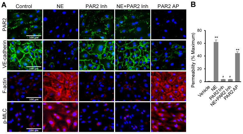
Figure 2.
NE regulated VE-cadherin, actomyosin cytoskeleton, and permeability in hECs through activating protease-activated receptor 2 (PAR2). (A) Immunofluorescence images of PAR2 (green), VE-cadherin (green), F-actin (red), or p-MLC (pSer19, red) and nuclei counterstaining DAPI (blue). hEC cells were treated with NE (20 nM) or PAR2 agonist (PAR2-AP, 7.5 μM) in the presence or absence of PAR2 inhibitor (1 μM) for 16 h. Images were taken at 20x using a fluorescence microscope; scale bar: 100 μm. (B) Graph showing percentage permeability of endothelial monolayer treated with 20 nM NE, 7.5 μM PAR2 agonist, and 1 μM PAR2 inhibitor. Permeability assay was carried out with Lucifer Yellow dye. All data are expressed as mean ± SD of three independent experiments. ** p < 0.01 vs. vehicle control group, # p < 0.05 vs. NE group. NE: neutrophil elastase, PAR2 Inh: protease-activated receptor 2 inhibitor (I191), PAR2 AP: protease-activated receptor 2 agonist (PAR2 (I-6) amide trifluroacetate salt). See also Figures S2–S4.

