Figure 4.
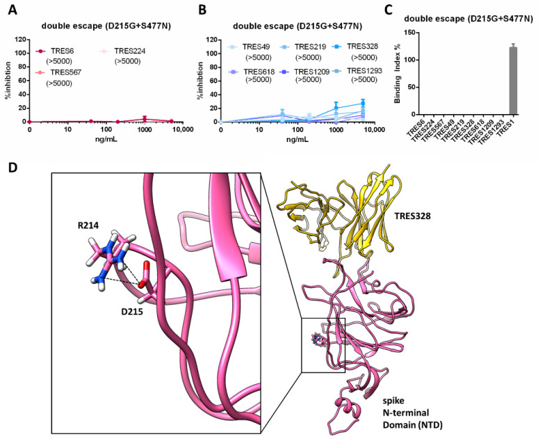
Characterization of S of the TRES6+TRES328 double escape variant. (A,B) Percent inhibition of lentiviral vector particles pseudotyped with SARS-CoV-2 S carrying the D215G and S477N mutations. IC50s of RBD (red) and NTD (blue) cluster antibodies are shown in ng/mL in the brackets. Neutralization and binding of the D614G wild-type spike protein is given in Figure 5A–C. (C) Binding of the RBD (red) and NTD (blue) cluster antibodies and an S2 binding antibody (grey) to S with the D215G and S477N mutations. (D) Structural modeling of TRES328 (yellow) in complex with the NTD of wild-type spike protein (pink). The NTD residues R214 and D215 are shown as sticks to illustrate that D215 is not located within the TRES328 interface and to highlight the close proximity of the R214 and D215 side chains, which form in some structures a salt bridge as strong electrostatic and stabilizing interaction (black dashed lines). The mutation to glycine (D215G) destroys this salt bridge interaction with R214, probably resulting in an increased flexibility of the arginine side chain, which could thereby possibly lead to a conformational change of the NTD.

