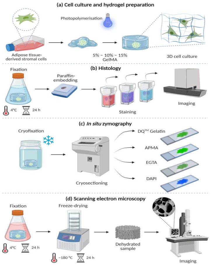
Figure 1.
Methods: (a) Culture of adipose tissue-derived stromal cells (ASCs) and loading in 5%, 10%, and 15% gelatine methacryloyl (GelMA) hydrogels. (b) Histology stains after 4% formalin-fixed and paraffin-embedding. Sections (4 µm) were stained with hematoxylin and eosin (H&E), picrosirius red (PSR) and phalloidin/DAPI stains to observe the cellular morphology and distribution within the GelMA hydrogels. (c) In situ zymography to detect matrix metalloproteases (MMPs). Hydrogels were cryofixed (liquid nitrogen), cryosectioned (4 µm), and exposed to four different conditions: DQTM Gelatin, DQTM Gelatin/APMA (MMP activator) DQTM Gelatin/EGTA (MMP inhibitor) and DAPI alone. Cell-free material was also stained. (d) Scanning electron microscopy (SEM) of GelMA hydrogels after fixation with 2% paraformaldehyde and 2% glutaraldehyde followed by freeze-drying and visualised at 1000× magnification. Created with biorender.com (accessed on 18 July 2022).

