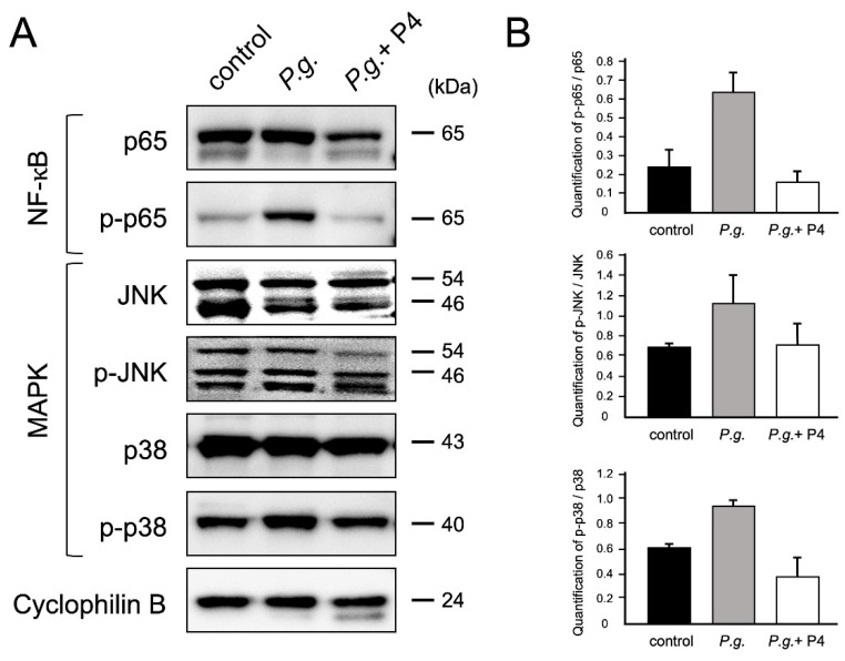
Figure 2.
P4 inhibited P.g.-induced NF-kB and MAPK phosphorylation (A) Western blot analysis was performed to detect the phosphorylation levels of NF-κB and MAPK signaling. Lane 1: control mice; Lane 2: P.g. mice; Lane 3: P.g. + P4 mice. (B) Densitometric data of protein analysis. The phosphorylated/total protein ratio was then quantified. Total and phosphorylated proteins were normalized to cyclophilin B in each group (control, P.g., and P.g. + P4). Experiments were performed using three independent tissue samples. Values represent the mean ± SD (n = 3).

