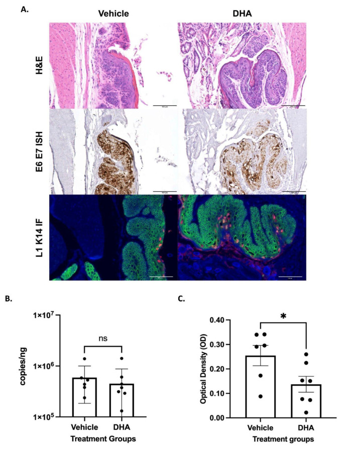
Figure 2.
NSG mice infected with MmuPV1 in the anal canal began treatment 20 weeks post infection with either topical DHA (N = 8) or vehicle (N = 6). (A) Representative images from MmuPV1-infected NSG mouse samples after completing treatment and stained with hematoxylin and eosin, RNAscope ISH (RNAscope® Probe: MusPV-E6-E7), and immunofluorescence staining for L1 (red) and K14 (green). All scale bars equal 100 μm. (B) Viral load analysis from NSG mice infected with MmuPV1 in the anus. Anal lavages were performed on vehicle mice (N = 6) and DHA-treated mice (N = 8) prior to sacrifice. DNA was extracted from the lavages, and qPCR was run for the E2 gene and normalized against 18s RNA. Treatment groups were compared via Mann–Whitney test. (C) RNAscope ISH anal tissue analysis from MmuPV1-infected NSG mice. Groups, vehicle mice (N = 6), and DHA-treated mice (N = 7) were compared via an unpaired t-test. Significance was assigned as not significant (ns), * p < 0.05.

